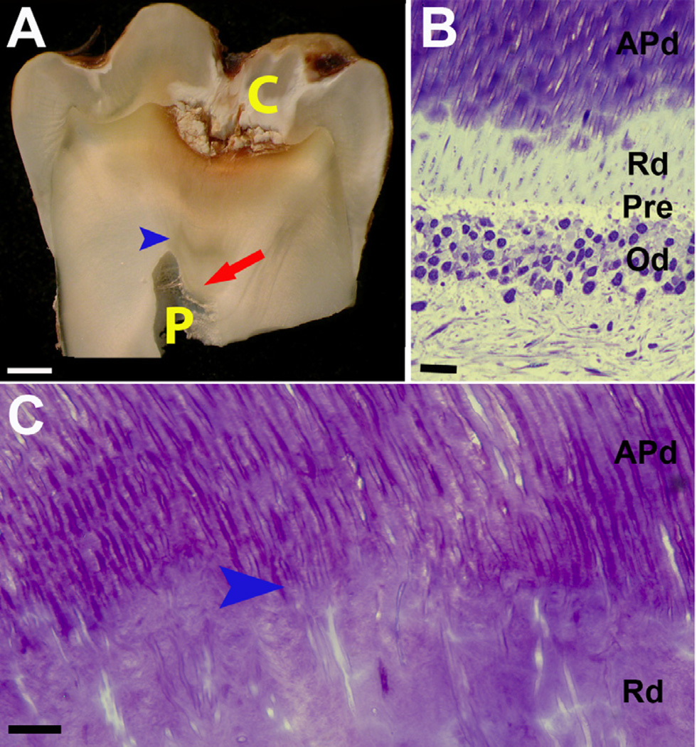
Figure 1. Representative images demonstrating the localized response of reactionary dentin (Rd) formation.
(A) In this carious tooth, Rd (red arrow) is deposited beneath the carious lesion (C) and affected physiological dentin (APd) with an obvious boundary between APd and Rd (blue arrow head). The pulp (P) has been reduced by deposition of Rd. (B) Toluidine blue stain of longitudinal section from an early stage of Rd formation showing the interface between odontoblasts (Od) and dentin. The limited amount of Rd is located inferior to APd but superior to pre-dentin (Pre) and the odontoblasts (Od). Scale bar 10 µm. (C) High magnification of Toluidine blue of longitudinal section of dentin from carious tooth presents the structure of Rd relative to APd. The varying course and wide to narrow lumen observed in Rd suggestive of spiral conformation. Scale bar 10 µm.

