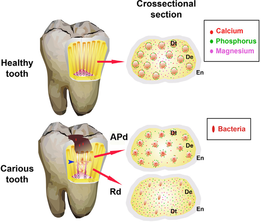
Figure 11. Diagrammatic representation of tooth structure affected by the carious Process.
Healthy tooth. Cross-section shows the enamel (En), dentin (De) and dentinal tubules (Dt) with mineral content. A highly mineralised calcium and phosphorus annular ring surrounding each Dt is observed with dispersion of magnesium in peritubular and inter-tubular dentin. Carious tooth. When bacterial invasion occurs calcium is lost from peritubular dentin and forms an intra-tubular plug. The phosphorus annular ring persists in affected physiological dentin (APd), but magnesium decreases. Reactionary dentin (Rd) formed in response to carious stimulation displays lower numbers of tubules and odontoblast processes. The cross-section shows reduction of diameter of tubules with scattered distribution of all elements but higher intensity of phosphorus and magnesium compared to dentin from healthy teeth and APd. The modified helical tubular structure in Rd (blue arrow) helps prevent the invasion of bacteria and potentially, acts as a shock absorber to compensate for the loss of upper tooth structure due to the carious lesion.

