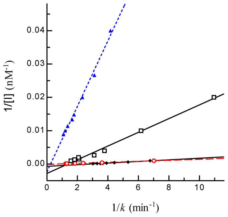
Fig. 5. Determination of Kd and k2 values of four inhibitors of A. gambiae AChE2.

Aliquots of the diluted enzyme (10 μl, 4.35 ng/μl) were individually added to 80 μl ATC-DTNB premixed with 10 μl carbaryl (□ ── □), eserine (▲ --- ▲), malaoxon (○ - - ○), or paraoxon (◆ ── ◆) at different concentrations. Absorbance at 405 nm was monitored immediately on the microplate reader at fifteen-second intervals for 5 min and the readings were used to derive k’s by curve fitting [A = A∞ (1−e−kt)]. Then, 1/k and 1/[I] values were plotted and analyzed by linear regression as described in Section 2.7 ([S] = 600 μM, KM = 5.075 μM, α = 0.992).
