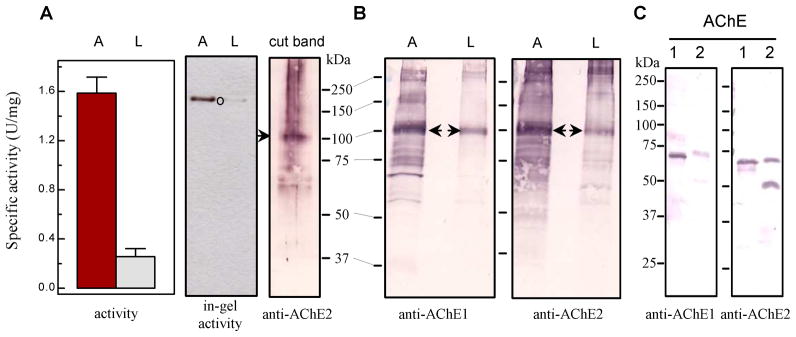
Fig. 8. Separation of proteins in larval and adult head extracts by native PAGE, in-gel activity assay, and immunoblot analysis.

(A) Left panel: Activity assay was performed using adult (A) and larval (L) head extracts (3.25 μg total protein in 20 μl buffer) mixed with 80 μl ATC-DTNB substrate solution (Section 2.6); Middle panel: in-gel AChE activity staining; Right panel (cut band): activity band (marked by ○) was cut from the “A” lane and subjected to reducing SDS-PAGE and immunoblot analysis using diluted AChE2 antiserum as the primary antibody. (B) Adult and larval head extracts (7.3 μg protein per lane) were separated by 7.5% SDS-PAGE, transferred onto nitrocellulose membrane, and incubated with diluted AChE1 (left panel) and AChE2 (right panel). The 100 kDa band (marked by arrow) was identified by mass spectrometric analysis as A. gambiae AChE1. Sizes and positions of Mr markers are indicated. (C) Cross-reactivity of the AChE1 and AChE2 antisera: Purified catalytic domains of A. gambiae AChE1 (lane 1, 50 ng) and AChE2 (lane 2, 50 ng) were separated by SDS-PAGE under reducing condition and electrotransferred to nitrocellulose membrane for immunoblot analysis using the diluted AChE1 (left panel) or AChE2 (right panel) antiserum as the primary antibody.
