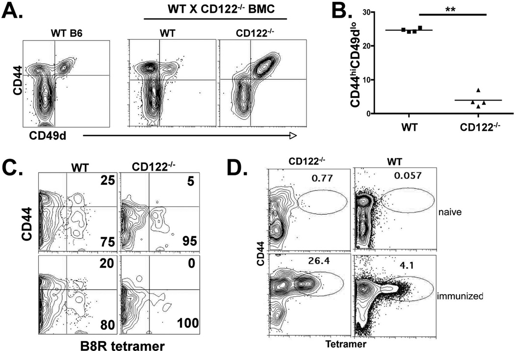
Figure 3.
VM cell development is CD122-dependent. WT × CD122−/− bone marrow chimeras were made as previously described and as in the Material Methods. Twelve weeks after reconstitution, spleen CD8+ T cells were analyzed for CD44 and CD49d expression. B) Percent of VM phenotype cells out of total CD8+ T cells from each background, WT or CD122−/− in the bone marrow chimeric mice shown in A. Each data point represents a separate chimeric host. Data are representative of 3 independent mixed chimera experiments, each with 3–5 chimeras. C) B8R specific T cells were isolated from the chimeric mice using magnetic enrichment of tetramer stained cells as described previously (15) and in the Materials and Methods. The column bound CD8+ cells were gated into CD45.1 (WT) and CD45.2 (CD122−/−) backgrounds and analyzed for CD44 expression and tetramer staining. Two representative mice are shown (top and bottom plots). D) Peripheral blood from WT×CD122−/− chimera’s before and after immunization with B8R peptide in conjunction with polyIC and antiCD40 as previously described (ref). Data shown are gated on all B220− CD8+CD45.1+ (WT) and B220−CD8+CD45.2+(CD122−/−) events. Data shown are representative of 4 immunized chimeric mice.

