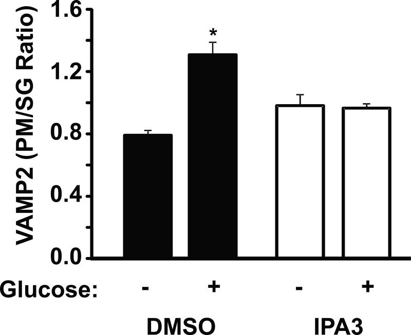
Figure 6. PAK1 is required for glucose-stimulated insulin granule accumulation at the β cell plasma membrane.
MIN6 cells were preincubated for 2 h in MKRBB and either vehicle (DMSO) or 30 μM IPA3 was added 10 min prior to stimulation with 20 mM glucose for 20 min. Cells were harvested and subjected to subcellular fractionation. Isolated plasma membrane (PM) and secretory granule (SG) fractions were analyzed by SDS-PAGE and immunoblotting (IB) for the granule v-SNARE and marker protein VAMP2. Optical density quantitation of VAMP2 in the PM versus the SG for each of three independent sets of fractions yielded the VAMP2 PM/SG ratio, represented in the bar graph as the average ± S.E.; *P<0.05, versus unstimulated vehicle fractions).

