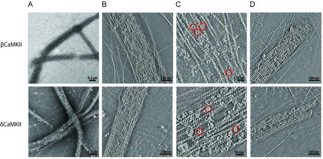
Figure 2. Structural analysis of actin bundles formed in the presence of the β and δ isoforms of CaMKII.
A) Left panels illustrate representative 2D projections of low power electron micrographs of negative stained F-actin in the presence of β (upper) and δ (lower) isoforms (n > 5). B) Panels show ~10 nm thick slices of two tomographic reconstructions to illustrate packing of β and δ CaMKII holoenzyme molecules within bundles. C) Several CaMKII holoenzyme molecules are highlighted by red circles in a zoomed in region of a representative slice from the same reconstructions shown in Panel B. D) The right panels show ~ 10 nm slices through two different tomographic reconstructions that illustrate examples of blunt ended bundles formed with β or δ CaMKII.

