
Figure 3.
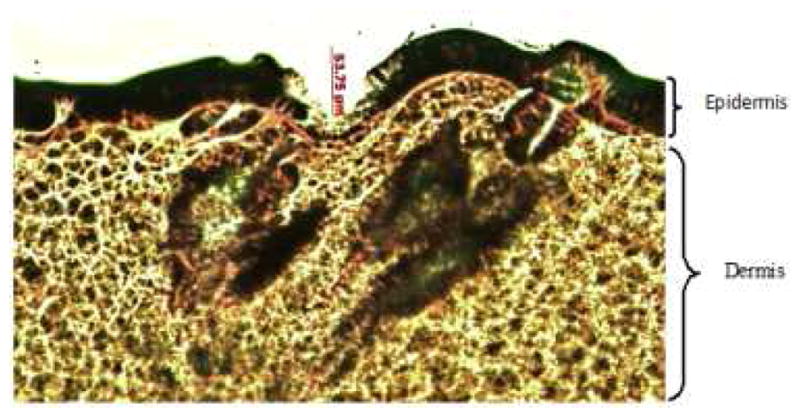
Histological section of skin treated with microneedles and stained with hematoxylin and eosin stain shown at 10x magnification. The epidermis and dermal layers were shown clearly in this picture.
Official websites use .gov
A
.gov website belongs to an official
government organization in the United States.
Secure .gov websites use HTTPS
A lock (
) or https:// means you've safely
connected to the .gov website. Share sensitive
information only on official, secure websites.
Histological section of skin treated with microneedles and stained with hematoxylin and eosin stain shown at 10x magnification. The epidermis and dermal layers were shown clearly in this picture.