
Figure 2. Cartoon outlining the procedure for preparation of LipoCEST microcapsules.
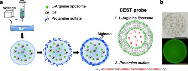
a, A mixture of cells, alginate and L-arginine liposomes is passed through a needle using a nanoinjector pump. The single-layered charged alginate droplets are collected, washed and resuspended in a crosslinker solution (protamine sulfate, PS), followed by a coating with a second layer of alginate. The L-arginine (R) moieties (red) entrapped in liposomes (1.) and in PS (2.) have exchangeable protons that provide CEST contrast. b, Phase contrast image (top) and fluorescent image (bottom; with NBD-PE labeled liposomes) shows a uniform distribution of cells and liposomes within the microcapsule. Scale bar = 200 μm.