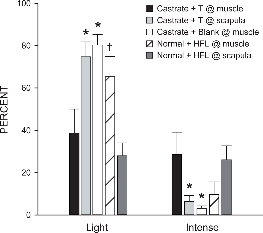
Figure 1.
Histogram of the number of lightly and intensely immunostained SNB motoneuron somata after immunolabeling for BDNF in castrated males with a testosterone (T) implant placed at the target muscle (black bars), castrated males with a T implant placed interscapularly (lightly shaded bars), castrated males with a blank implant placed at the target muscle (open bars), gonadally intact males with a hydroxyflutamide (HFL) implant placed interscapularly (hatched bars) and gonadally intact males with a hydroxyflutamide implant placed at the target muscle (darkly shaded bars). Bar heights represent means ± SEM. * Significantly different from castrated males with a T implant placed at the target muscle; † Significantly different from gonadally intact males with a hydroxyflutamide implant placed interscapularly. (Verhovshek et al., 2010a)

