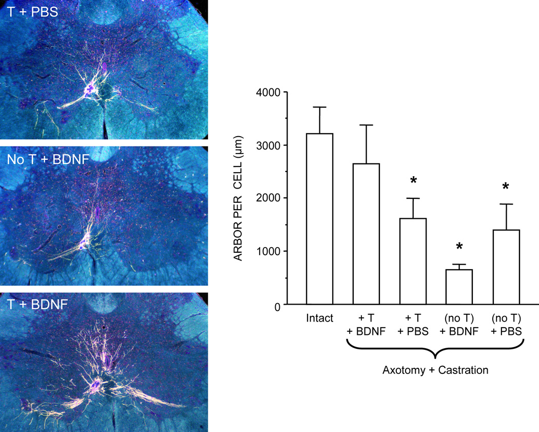
Figure 4.
(Left) Dark-field digital images of transverse sections through the lumbar spinal cord showing BHRP labeling of SNB dendrites in axotomized and castrated males treated with testosterone (T) and phosphate buffered saline (PBS) (top), BDNF alone (middle), or T and BDNF (bottom). (Right) Dendritic length per labeled motoneuron in intact males and axotomized and castrated males with T and BDNF (T+BDNF), T alone (T+PBS), BDNF alone (no T+BDNF), and PBS alone (no T+PBS) applied to the cut SNB axons. Bar heights represent means ± SEM. * Significantly different from intact males. (Adapted from Yang et al., 2004)

