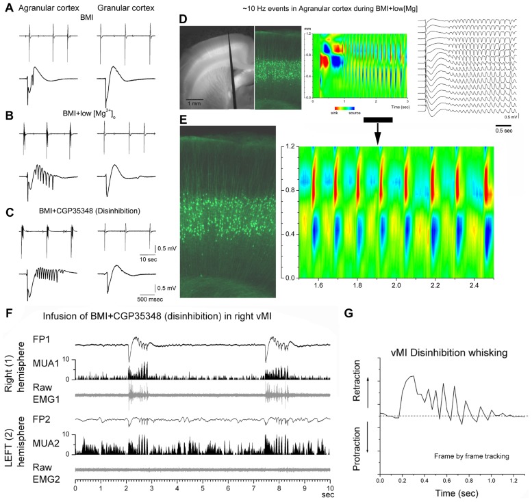
FIGURE 2.
7–14 Hz oscillations are caused by low [Mg2+]o buffer or disinhibition in the motor cortex but not in the somatosensory cortex. (A–C) Field potential (FP) recordings in neocortical slices showing typical spontaneous events recorded in motor (agranular) cortex (left panels) and in somatosensory (granular) cortex (right panels) during block of GABAA receptors with bicuculline (BMI) (A), subsequent lowering of [Mg2+]o (B), or subsequent block of GABAB receptors (C). Note the occurrence of 7–14 Hz oscillations only in agranular cortex. In each panel, the upper trace has a long time scale and the lower trace shows a close-up of a spontaneous discharge (Castro-Alamancos and Tawara-Hirata, 2007). (D) Recording from a fluorescent slice of an Thy1-eYFP mouse using a 16-channel silicon probe. The left panel shows a light/fluorescent image of the probe position in the motor cortex during the experiment. The middle left panel show a fluorescent image taken at the probe location revealing the layer V fluorescent cells. The current source density (CSD) and FP plots show a 7–14 Hz oscillation event. The CSD and fluorescent image are scaled to match the cortical layers location with the location of the current flow (Castro-Alamancos and Tawara-Hirata, 2007). (E) Close-up of the 7–14 Hz event shown in (D). (F) 7–14 Hz oscillations caused by disinhibition in the whisker motor cortex (vMI) of a freely behaving rat. Bilateral field potential (FP) and multi-unit (MUA) activity recorded from the vMI during disinhibition caused by application of BMI + CGP (0.1 + 2 mM, 0.2 μl volume) into the right vMI. The corresponding raw electromyography (EMG) activity measured from the contralateral whisker pad is also displayed below the FP and MUA. 1 refers to the infused hemisphere and the related (contralateral) whisker pad, 2 refers to the non-infused hemisphere and the related whisker pad (Castro-Alamancos, 2006). (G) 7–14 Hz oscillations of vMI caused by disinhibition produce cycles of vibrissa retractions. Frame by frame tracking of a vibrissa movement caused by vMI disinhibition. Units in the y-axis are relative pixels (see Castro-Alamancos, 2006).

