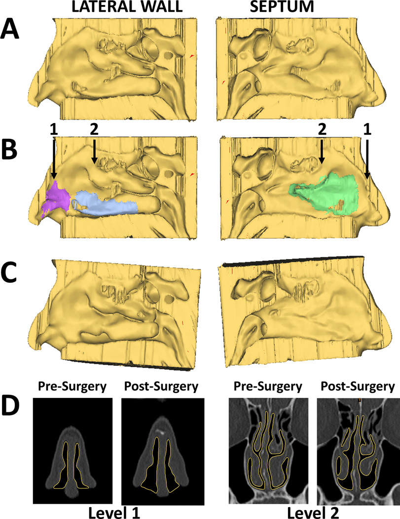
Figure 1.
(A) Reconstructions of right lateral wall and right side of septum from pre-surgery CT scan. (B) Same views with surgical sites marked. Purple=nasal valve repair; blue=right inferior turbinate reduction; green=septoplasty. Arrows indicate coronal levels shown in (D). (C) Reconstructions of right lateral wall and right septum from post-surgery CT scan, coregistered with pre-surgery scan. (D) Coronal views from pre- and post-surgery CT scans. Yellow outline highlights border between soft tissue and nasal air space.

