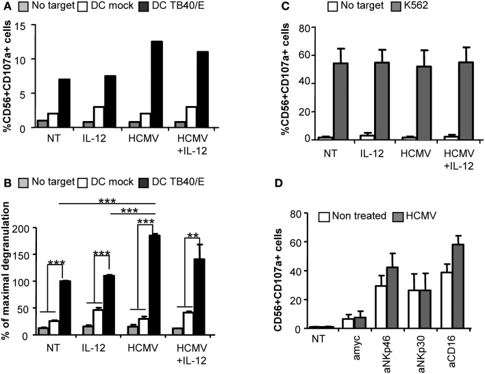
Figure 3.
Direct HCMV recognition enhances NK cell degranulation against HCMV-infected moDC. Freshly purified NK cells were incubated alone (NT), or with IL-12 and/or HCMV TB40/E. After 24 h, cells were washed and the degranulation of NK cells was assessed after 5 h incubation with mock or HCMV TB40/E infected moDC (A,B), and K562 (C) (E:T, 4:1). NK cell degranulation was monitored by CD107a staining analyzed by flow cytometry. (A) Bar graphs represent the percent of NK cells positive for CD107a in the absence of target cell (gray bars), in front of mock moDC (empty bars), or HCMV-infected moDC (black bars) from one representative donor. (B) Bar graph displaying the mean ± SEM of the normalized degranulation. Data in each experiment were normalized to the response of non-treated NK cells incubated with HCMV-infected DC (100%). Statistical analysis by the Student t-test (**p < 0.01; ***p < 0.0001) (C) Bar graph showing the mean ± SEM of the percent in degranulation to K562 of three donors tested (D) Bar graph displaying the mean ± SEM frequency of CD56+CD107a+ of non-treated (empty bars) or HCMV treated (gray bars) NK cells responding to P815 cells coupled to mAbs specific for NKp46 (Bab281), NKp30 (az20), CD16 (KD1), and anti-myc as control IgG in three independent experiments.

