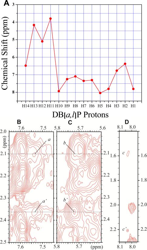
Figure 4.
(A) Plot of the chemical shifts of the DB[a,l]P aromatic ring protons. (B) - (D): Expanded contour plots of the NOESY spectrum (200ms mixing time) of the DB[a,l]P-dG lesion in the 11mer duplex in D2O aqueous buffer solution using a 500MHz spectrometer with a cryoprobe at 28 °C. (B) NOEs between the C7 base H6 proton and its own 2′, and 2″ sugar protons: a, C7(H6) – C7(H2′); a’, C7(H6) – C7(H2″). (C) NOEs between C7-H1′ and its own 2′ and 2″ sugar protons: b, C7(H1′) – C7(H2′); b’: C7(H1′) C7(H2″). (D) NOEs between the G6* base proton H8 and flanking base C5 sugar protons: c, G6*(H8) – C5(H2′); c’, G6*(H8) – C5(H2″).

