
Figure 2.
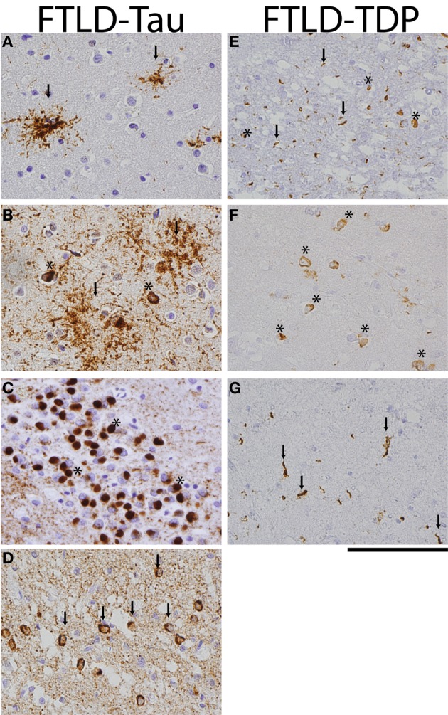
FTLD-Tau and FTLD-TDP histology. Photomicrographs of FTLD-tau (A–D) and FTLD-TDP (E–G) visualized with immunohistochemistry (PHF-1 and pTDP 409/410 for tau and TDP, respectively). (A) PSP frontal cortex with tau-positive tufted astrocytes (arrows), (B) CBD temporal cortex with diffuse astrocytic plaques (arrows) and neuronal tangles (asterisks), (C) Pick's disease with round tau-positive Pick bodies (asterisks) in the dentate nucleus of the hippocampus, (D) FTDP-17 case with p.P301L pathogenic mutation with tau-positive neuronal tangles (arrows) and diffuse neuropil threads in temporal cortex, (E) FTLD-TDP subtype A with cytoplasmic neuronal inclusions (asterisks) and short dystrophic neurites (arrows) in superficial layers of frontal cortex, (F) FTLD-TDP subtype B with prominent cytoplasmic inclusions (asterisks) in deep temporal cortical layer, and (G) long dystrophic neurites (arrows) in superficial layers of mid-frontal cortex of a patient with FTLD-TDP subtype C. Scale bar = 100 μm.