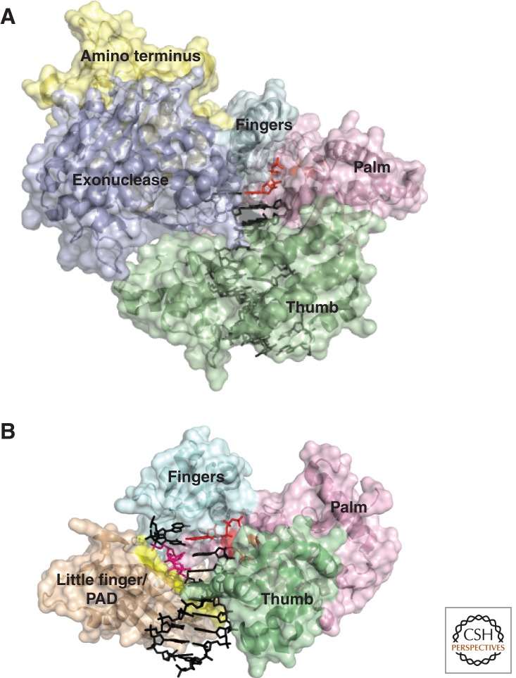
Figure 2.
Comparative anatomy of a replicative polymerase and a TLS polymerase. (A) S. cerevisiae polymerase δ, PDB 3IAY (Swan et al. 2009), a replicative B-family polymerase. The domains are shaded: palm, pink; thumb, green; fingers, cyan; exonuclease, purple. The DNA is in black, and the active site in the palm and incoming nucleotide triphosphate is in red. (B) H. sapiens polymerase η, PDB 3SI8 (Biertumpfel et al. 2010), a Y-family TLS polymerase with a T-T CPD in the +2 position. The domains in common with Pol δ are shaded the same. The little-finger domain/PAD is shaded in light brown. The DNA is in black, except the CPD, which is pink. The active site and incoming nucleotide triphosphate pairing with the first base after the CPD are in red. The β-strand splint in the little finger/PAD domain that constrains the CPD is highlighted in yellow.

