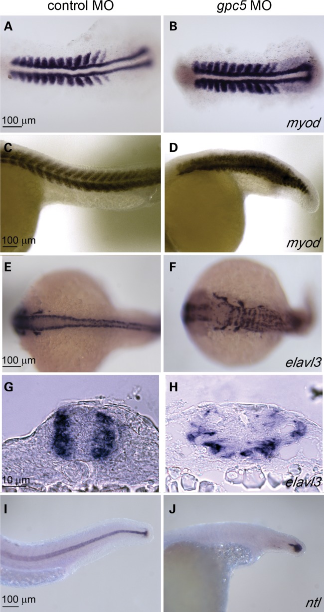
Figure 4.
gpc5 MO disrupt morphogenesis of the trunk in zebrafish embryos. Abnormal morphogenesis of the neural tube and surrounding tissue in gpc5 MO-injected embryos compared with controls, (A–J). Embryos fixed at 14 hpf (A and B) or 24 hpf (C–J) were stained for myod (A–D), a marker of somatic mesoderm; elavl3 (E–H), a marker of differentiated neurons; or ntl (I,J), a marker of the notochord. In comparison to (E) control MO-injected embryos, the hindbrain is broader and neurons appear disorganized in (F) gpc5 MO-injected embryos. This is highlighted in transverse trunk cross sections of (G) control and (H) gpc5 MO-injected embryos. In (A) control MO-injected embryos, somites exhibit a classic chevron shape, while in (B) gpc5 MO-injected embryos, myod expression is compressed. (I and J) In lateral view, the notohord is clear in (I) control MO-injected embryos, while it is absent in (J) gpc5 MO-injected embryos.

