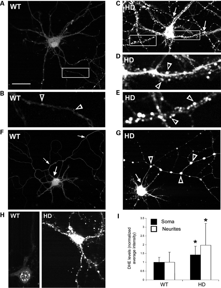
Figure 3.
Spines and axons of HD140Q/140Q primary cortical neurons have high levels of ROS as detected by DCF fluorescence. Shown are images of living neurons from WT (A, B, F) and HD140Q/140Q (C–E, G) cultures of embryonic cortex grown to 8 DIV. (A and C) Positive DCF-labeled WT (A) and HD140Q/140Q (C) neurons. The boxed areas are shown at higher magnification in B for the WT neuron and in D and E for the HD140Q/140Q neuron. Varicosities are prominent in HD140Q/140Q neurites (C and G, arrows in C and open arrowheads in G) and not WT neurites (A and F). Scale bar in (A) represents 25 μm. (D and E) Small brightly DCF fluorescent spines (open arrowheads) occur along the neurites of the HD140Q/140Q neuron shown in (C). (F and G) The axon is recognized as the only neurite that emerges from the cell body (arrow) and form branches that extend beyond the field of the other neurites. Note the large DCF fluorescent varicosities (open arrowheads) in the axon of the HD140Q/140Q neuron. (H) Cortical neurons with DHE fluorescence. Live cell images of WT and HD140Q/140Q primary cortical neurons at 8 DIV show fluorescence from DHE which preferentially reacts with superoxide. Note that the DHE is retained in abnormal swellings of HD140Q/140Q neurons. (I) Bar graph shows densitometry results for DHE fluorescent neurons. Student's t-test *P < 0.01, n = 19 WT and 24 HD140Q/140Q neurons.

