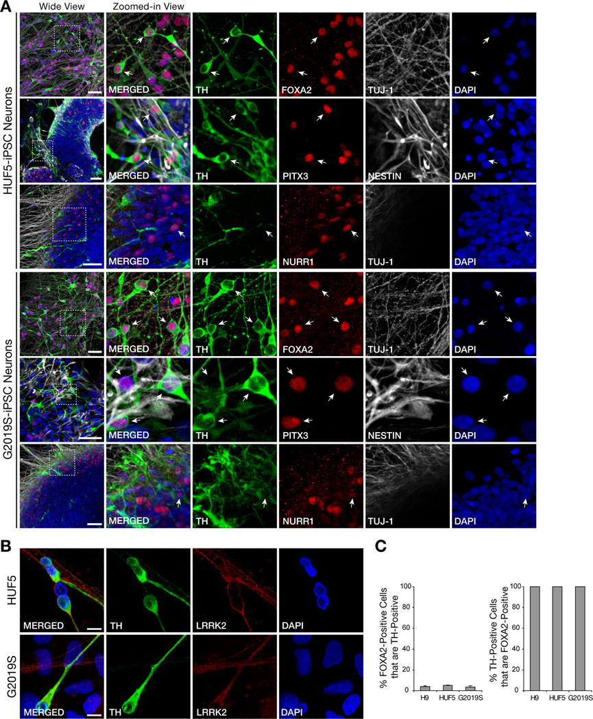
Figure 2. Directed Differentiation of iPSCs into Midbrain Dopaminergic (mDA) Neurons.
(A) Expression of midbrain dopaminergic neurons markers, tyrosine hydroxylase (TH), FOXA2, PITX3, and NURR1, in iPSC-derived 30- to 35-day neurons. Note the co-localization of FOXA, PITX3 and NURR1 with TH, characteristic of mDA neurons (arrows). NESTIN and TUJ1 positive immunoreactivity also show the presence of neurons. Scale bar = 50 µm.
(B) Expression of LRRK2 proteins in dopaminergic neurons. Scale bar = 10 µm.
(C) Quantification of the yield of mDA, based on TH and FOXA2 colocalization, in 35-day neurons derived from H9, HUF5, and G2019S lines.
See also Figure S2 for more characterization of iPSC-derived neurons.

