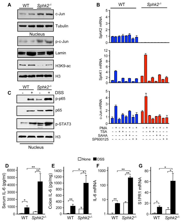
Figure 3. Severity of colitis in Sphk2−/− mice correlates with upregulation of SphK1, S1PR1, and IL-6, and activation of NF-κB and STAT3.
(A) Colonic lysates or nuclear extracts from Sphk2−/− mice and WT littermates were analyzed by immunoblotting with the indicated antibodies.
(B) WT and Sphk2−/− MEFs were pretreated without or with SAHA, TSA, or SP600125, stimulated without or with PMA for 3 hr, and mRNA levels were determined by QPCR.
(C–G) Colitis was induced with 5% DSS. Equal amounts of colonic nuclear extracts were analyzed by western blotting with the indicated antibodies (C). IL-6 in serum (D) and secreted from colon (E) was measured by ELISA. Expression of IL-6 (F) and S1PR1 (G) was determined in colons by QPCR.
Data are means ± SD. *p<0.05, **p<0.005.
See also Figure S2.

