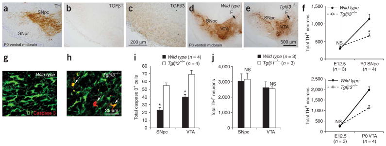
Figure 8.

Loss of TGFβ3, but not TGFβ1, leads to DA neuron deficiencies similar to those in Hipk2−/− mutants. (a–c) Immunohistochemistry shows that TGFβ1 is undetectable in the ventral mesencephalon at P0 (b). In contrast, a relatively high concentration of TGFβ3(c) is present in regions adjacent to DA neurons in the SNpc (a). (d,e) Reduction of DA neuron density is present in the SNpc and VTA of Tgfβ3−/− mutants at P0. Scale bar, 500 μm. (f) Quantification of DA neurons in wild-type and Tgfβ3−/− mutants shows no difference at E12.5, but does show a 50% reduction in both the SNpc and VTA at P0. * indicates two-tailed Student’s t-test, P = 0.018 for SNpc and P = 0.002 for VTA, n = 3 for E12.5 and n = 4 for P0. (g,h) Increased apoptotic cell death is present in the SNpc and VTA in Tgfβ3−/− mutants at P0. Confocal images of double immunofluorescence using activated caspase 3 (red) and TH (green). Arrows indicate double positive cells. Scale bar, 25 μm. (i) Quantification of DA neurons and activated caspase 3–positive cells in the SNpc and VTA of Tgfβ3−/− mutants shows deficits similar to those in Hipk2−/− at the same stage. (j) No detectable loss of DA neurons is found in the SNpc or VTA of Tgfβ1−/− mutants at P28. NS indicates not significant, two-tailed Student’s t-test, n = 3 for each genotype.
