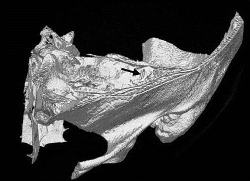
Figure 3.

The arrow indicates the dehiscence observed in this computed tomography image from reconstructions in the plane of the left superior canal. The patient is the 39-year-old male described in Fig. 2. (Reprinted with permission from Minor LB. Clinical manifestations of superior semicircular canal dehiscence. The Laryngoscope 2005;115:1717–1727.)
