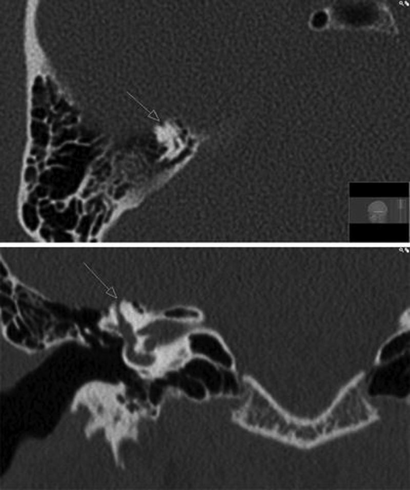
Figure 4.

Computed tomography scan of a superior semicircular canal dehiscence in the right ear. The axial view is displayed above the coronal view. (Reprinted with permission from Stimmer H, Hamann KF, Zeiter S, Naumann A, Rummeny EJ. Semicircular canal dehiscence in HR multislice computed tomography: distribution, frequency, and clinical relevance. Eur Arch Otorhinolaryngol 2012;269:475–480.)
