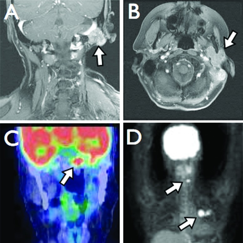
Figure 4.
(A) Coronal and (B) axial T1-weighted magnetic resonance imaging (MRI) with gadolinium demonstrating involvement of the left temporal bone, sigmoid sinus, and mandible. (C) Coronal and (D) three-dimensional positron emission tomography (3D PET) imaging reveals metastases at the clivus, cervical spine, and left lung.

