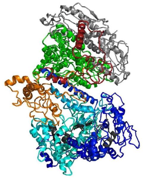
Figure 2.


Functional and structural domains of the polymerase protein. A. Linear organization of the polymerase gamma protein. The N-terminal domain (NTD) region is the mitochondrial targeting sequence and is located at the N-terminal of the protein. The thumb subdomains (Th) of the protein and are found between the exonuclease and linker region as well as within the polymerase region [63]. The exonuclease domain contains essential motifs I, II, and III for its activity. The polymerase domain contains subdomains comprising the thumb (Th), palm, and finger, which contain motifs A, B, and C, respectively. There are two regions that make up the palm subdomain within the polymerase domain. The motifs located within the polymerase domain are critical for polymerase activity [23, 57]. The small arrows indicate position of the numbered amino acid position that begin a subdomain. B. Three dimensional structure of the human DNA polymerase holoenzyme [59]. The color scheme of the polymerase gamma catalytic subunit is the same as in A (light blue for the polymerase domain, dark blue for the exonuclease region, red for the accessory interacting domain (AID) domain and orange for the intrinsic processivity (IP) domain. The conserved motifs in the exonuclease (I, II, and III) and in the polymerase (A, B, and C) are colored in charcoal. The two accessory subunits are colored in green, for the proximal subunit, and grey for the distal subunit.
