
Figure 4.
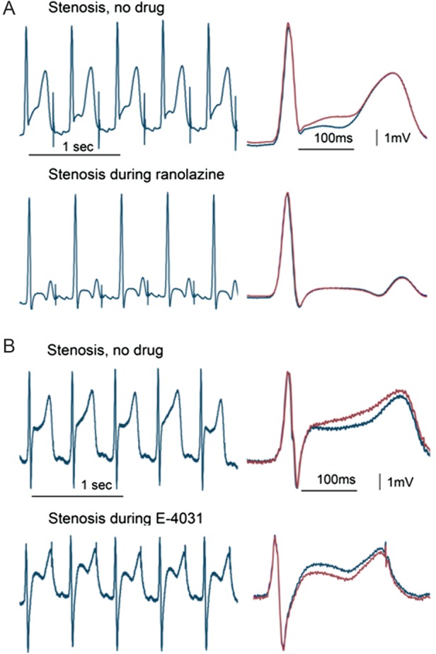
T-wave alternans (TWA) during left anterior descending coronary artery stenosis. The tracings with and without ranolazine (A) or E-4031 (B) are continuous (left panels) and with QRS-aligned superimposition (right panels). The stenosis-induced TWA was suppressed by ranolazine but not by E-4031. (Reprinted from ref.25 with permission from Heart Rhythm Society.)