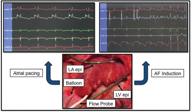
Figure 5.
Experimental setup. Upper left panel: left atrial (LA) and left ventricular (LV) epicardial electrocardiograms obtained prior to balloon occlusion of the left circumflex (LCx) coronary artery to reduce flow by 75% during atrial pacing at 150 beats/min. Upper right panel: Induction of atrial fibrillation (AF) by a 6-mA S2 test stimulus following the last S1 pacing stimulus. Note visible T-wave heterogeneity compared to the uniform pattern observed in the upper left panel. Lower panel: Hydraulic balloon occluder positioned around the proximal LCx coronary artery upstream of the Doppler flow probe. Electrode catheters are affixed to the left atrial appendage and left ventricular epicardium within the atrial and ventricular regions supplied by the LCx. (Reprinted from ref. 47 with permission from Heart Rhythm Society.)

