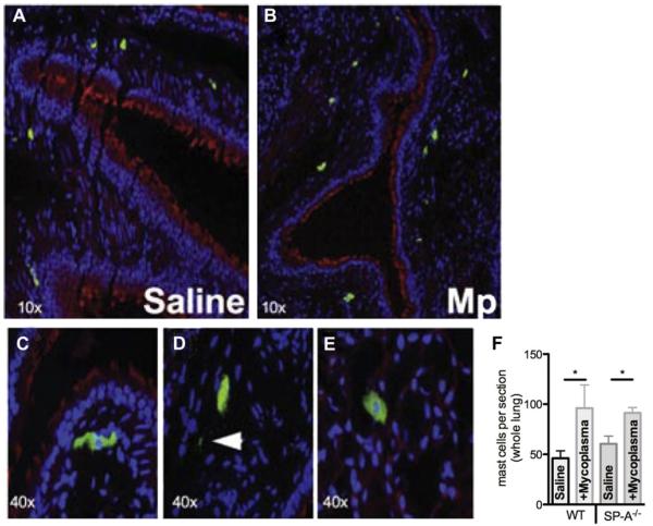
FIG 2.
MCs in Mycoplasma pneumoniae (Mp)–infected lung tissue. Mice were instilled with saline (A) or M pneumoniae (B-E) and lungs were analyzed by means of immunohistochemistry 3 days after infection for MCs. Fig 2, C, MCs adjacent to the large airway. Fig 2, D, Extracellular MC granules (arrow). Fig 2, E, MCs in the lung parenchyma. F, Total number of tissue MCs assessed in WT versus SP-A−/− lungs (n = 5 each). *P < .05.

