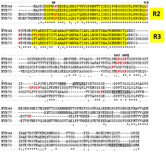
Figure 2. Protein alignment of MYB subfamily S22.
The R2R3 repeats of the DNA-binding domain are indicated by yellow boxes. Ser-Pro dipeptides matching the minimal MAPK phosphorylation consensus motif (red) or putative MAPK docking sites (R/K x2-6 I/L×I/L) (grey) are highlighted. MYB44 Ser142 and Ser145 are indicated. *conserved residues.

