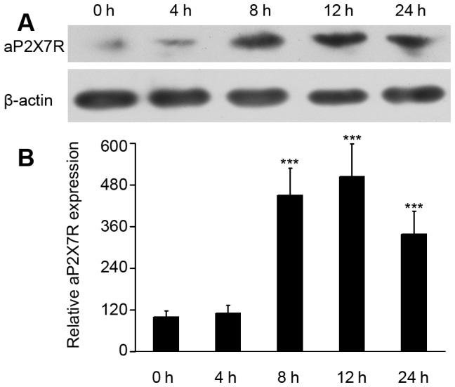
Figure 5. Western blot analysis of aP2X7R protein expression in macrophages upon L. anguillarum treatment.

(A) Protein collected from L. anguillarum-infected macrophages at 0, 4, 8, 12 and 24 hpi was assayed by Western blotting using an antiserum specific to aP2X7R. (B) Histogram displaying the changes in relative band intensity of aP2X7R protein in L. anguillarum-infected macrophages at 0, 4, 8, 12, and 24 h. Data are representative of three independent experiments. ***P<0.001.
