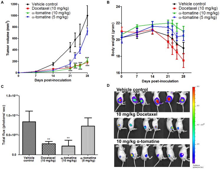
Figure 4. Anti-tumor activity of α-tomatine against subcutaneous PC-3 cell tumors.
Luciferase expressing PC-3 cell xenograft tumors established in male nude mice (n = 8 per treatment group) for 1 week were treated thrice weekly for 3 weeks with vehicle, docetaxel (10 mg/kg) or α-tomatine (5 or 10 mg/kg). (A) Graph of tumor volume in each treatment group versus the number of days after initial injection of PC-3 cells. (B) Graph of mean body weight for each treatment group versus the number of days after initial injection of PC-3 cells. (C) Bioluminescence intensities emitted from PC-3 cell xenograft tumors at the end of the experiment for each treatment group. (D) Bioluminescence images of PC-3 subcutaneous xenografts. The first row shows the vehicle control group; middle row shows the docetaxel treatment group; bottom row shows the 10 mg/kg α-tomatine treatment group. Each bar or point represents the mean ± SEM of data (n = 8).* P<0.05, ** P<0.01, *** P<0.001 vs vehicle control.

