
Figure 2. T. gondii tachyzoites infect human retinal glial cells in preference to neurons.
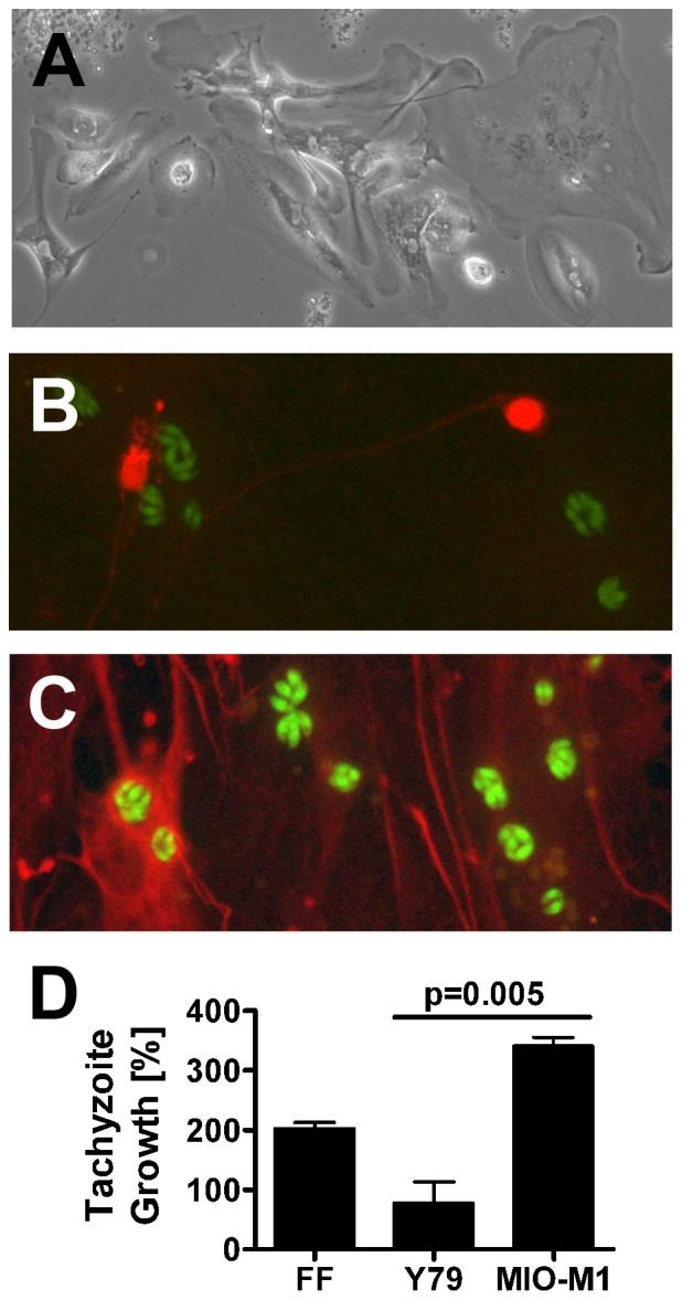
(A). Immediately prior to infection, dissociated human retinal cultures presented a layer of glial cells with neurons positioned above. Original magnification: 100X. (B and C). Expression of (B) neuron specific enolase (NSE) (red), as detected by rabbit polyclonal anti-human NSE antibody and Alexa Fluor 594-conjugated donkey anti-rabbit immunoglobulin (Ig)G antibody and (C) glial fibrillary acidic protein (GFAP) (red), as detected by sheep polyclonal anti-human GFAP antibody and Alexa Fluor 594-conjugated donkey anti-sheep IgG antibody. T. gondii tachyzoites express YFP (green). Original magnification: 630X. Negative control cultures showed no positive staining. (D). Graph showing percentage growth of tachyzoites in Y79 human retinoblastoma cells and MIO-M1 human Müller glial cells, plus positive control human foreskin fibroblasts (FF), over a 24-hour period. n = 7–8 wells/condition. Columns = mean. Error bars = standard error of mean. Representative of two independent experiments.