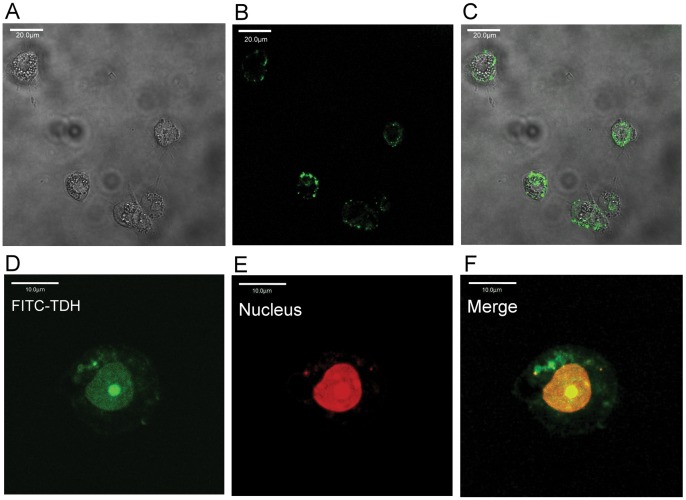
Figure 4. Subcellular localization of Gh-rTDH.
Liver cells were treated with 10 µg/ml Gh-rTDH-FITC for 20 (A–C) or 40 (D–F) min at 26°C and were then observed by confocal microscopy. (A) The liver cells were observed without a FITC filter, (B) with a FITC filter, (C) and with A and B merged, confirming that Gh-rTDH-FITC (green) could bind around the liver cell margins. (D) Gh-rTDH-FITC (green) was taken up by the nuclei of liver cells. (E) Nuclei stained with PI (red). (F) The merge of D and E confirmed that Gh-rTDH-FITC was located in the nuclei of liver cells.

