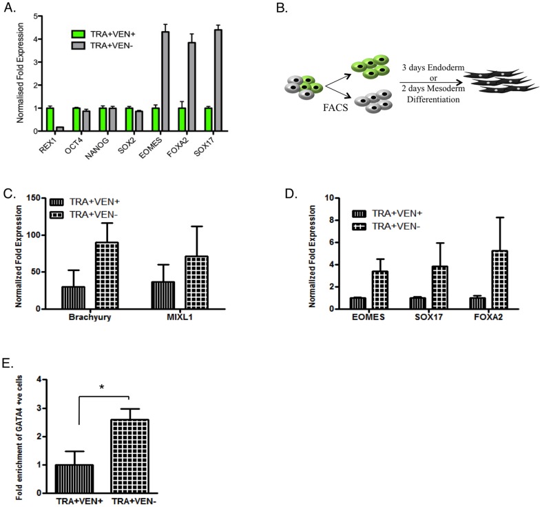
Figure 6. Loss of REX1 within the pluripotent population primes cells for differentiation.
A) QRT-PCR analysis of gene transcript expression in FACS separated TRA+VEN+ and TRA+VEN− populations. The TRA+VEN− fraction is normalised relative to the TRA+VEN+ population = 1. B) Schematic showing the differentiation treatment of hESCs C & D) QRT-PCR data showing the expression of mesoderm (C) and endoderm (D) lineage associated markers after the TRA+VEN+ and TRA+VEN− fractions were subject to 2 or 3 days of differentiation in mesoderm or endoderm conditions, respectively. C) BRACHYURY and MIXL1 were used as mesoderm associated markers. Undifferentiated TRA+VEN+ population was used as a control. n = 2 D) EOMES, SOX17 and FOXA2 were used as endoderm specific markers, and gene expression was normalized to TRA+VEN+ day 3 differentiated cells. n = 3 E) Fold enrichment of the percentage of GATA4 positive endoderm cells generated from TRA+VEN− cells relative to those from TRA+VEN+ population after 3 days of treatment with Activin A and BMP4 in low serum media, as observed by immunocytochemistry.

