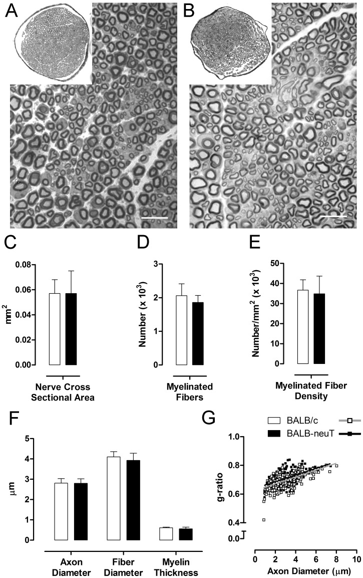
Figure 5. Morphological and stereological analysis of uninjured median nerves do not show detectable differences.
A, B: representative light micrographs of transverse sections of median nerves stained with toluidine blue of BALB/c and BALB-neuT mice respectively. In both groups myelinated axons have a normal morphological appearance. Bar = 10 µm. C, D, E, F: histograms showing the results of histomorphometric evaluations. No significant differences are detectable for all analyzed parameters. Values in the graphics are expressed as mean+SEM. G: Scatterplots displaying g-ratios of individual fibers in relation to respective axon diameter (obtained from more than 250 myelinated axons per group, 5 mice per genotype) are not different in BALB/c and BALB-neuT mice.

