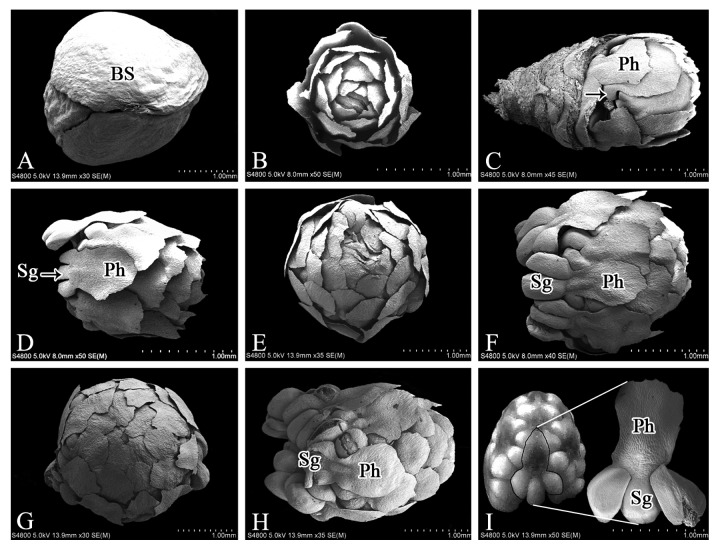
Figure 2. SEM images of male cones of M. glyptostroboides at different developmental stages. (A) In early September, the male cone was subtended by decussate bud-scales. (B) In mid-September, the spirally arranged microsporophylls underwent differentiation. (C) By late September, microsporangium primordia were visible (arrow). (D) In early October, the microsporophyll was composed of the phylloclade and microsporangia. (E) The microsporophylls enlarged and were tightly arranged around the main axis. (F–G) In November, the microsporangia and phylloclade had enlarged significantly; (H) In December, the enlarged microstrobilus grew slowly; (I) In early February, mature microsporophyll contained 2–3 microsporangia and an phylloclade. BS, bud scales; Ph, phylloclade; Sg, sporangia. Bars = 1 mm.

An official website of the United States government
Here's how you know
Official websites use .gov
A
.gov website belongs to an official
government organization in the United States.
Secure .gov websites use HTTPS
A lock (
) or https:// means you've safely
connected to the .gov website. Share sensitive
information only on official, secure websites.
