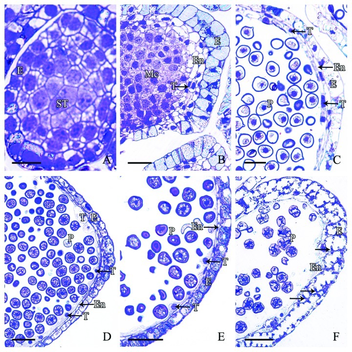
Figure 3. Stages of microsporangial wall development in M. glyptostroboides (A) In mid-September, the outermost epidermal cells differentiated and contained a large conspicuous nucleus. (B) In late September, the number of epidermal cells increased and the inner 2–3 layers of cells formed the early endothecium and tapetum layers, which showed no distinct differences. (C) In November, the linear-shaped tapetum and endothecium cell layers differentiated. (D–E) From early December to late January, the tapetum cells were well-developed and protruded inwards. (F) In mid-February, expansion and wall thickening of the epidermal cells occurred with protrusion inwards on cell walls, and the tapetum disintegrated. E, Epidermis; En, endothecium; Mc, mother cell; P, pollen; ST, sporogenous tissue; T, tapetum. Scale bars = 50 μm.

An official website of the United States government
Here's how you know
Official websites use .gov
A
.gov website belongs to an official
government organization in the United States.
Secure .gov websites use HTTPS
A lock (
) or https:// means you've safely
connected to the .gov website. Share sensitive
information only on official, secure websites.
