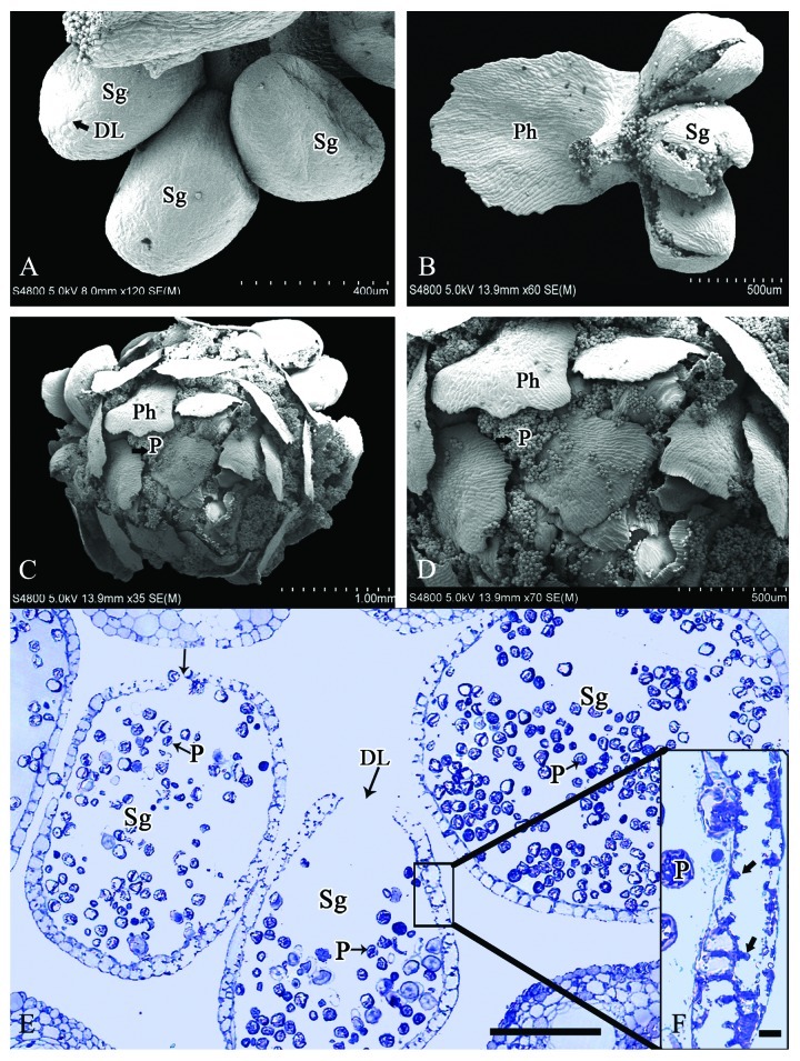
Figure 4. Microsporangia at the pollen dispersal stage in M. glyptostroboides. (A) Pollen grains are released through the dehiscence line, which formed early in microsporangium development in mid-October. (B) Microsporangial wall ruptured along the dehiscence line in February. (C–D) Mature pollen grains are dispersed spirally in the space between the phylloclades. (E) Transverse section of dehisced microsporangia in mid-February. (F) Thickening of microsporangial wall with many internal protrusions (arrows). DL, Dehiscence line; P, pollen; Ph, phylloclade; Sg, sporangia. Scale bars (A) = 400 μm, (B, D) = 500 μm, (C) = 1 mm, (E) = 100 μm, (F) = 10 μm.

An official website of the United States government
Here's how you know
Official websites use .gov
A
.gov website belongs to an official
government organization in the United States.
Secure .gov websites use HTTPS
A lock (
) or https:// means you've safely
connected to the .gov website. Share sensitive
information only on official, secure websites.
