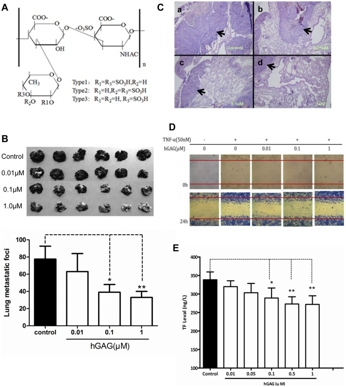
Figure 1. Effects of hGAG on aggressiveness of B16F10 tumor cells.
(A) Chemical structure of hGAG. (B) Representative metastatic nodules on lung tissues. B16F10 tumor cells were treated with hGAG at the indicated concentration for 24 hours/37°C and injected into C57BL/6J mice through the tail vein. After 23 days, mice were sacrificed and metastatic nodules on lung surface were photographed. Metastatic nodules were counted under a dissecting microscope. Values are expressed as the mean ± SD. hGAG treatment reduces in vivo metastatic capacity of B16F10 tumor cells in mice. (C) Representative HE staining of lung tissue sections. Paraffin-embedded formalin-fixed lung tissues from each group were prepared and the sections were H&E stained. Arrows indicated tumor cells on each section. (D) Wound healing. B16F10 monolayer cells at 90–95% confluence were serum starved for 24 h and then carefully wounded using sterilized pipette tips (t = 0 h). After removing detached cells, cells were incubated with medium, tumor necrosis factor (TNFα, final conc. 50 nM), or TNFα in combination with hGAG at the indicated concentration for 24 h at 37°C and photographed immediately (t = 24 h). (E) TF levels in the plasma from mice assessed by ELISA. hGAG-treated B16F10 tumor cells injected into mice produced reduced level of TF. Data was expressed as mean ± S.E. (3–5 independent experiments). *p<0.05, **p<0.01.

