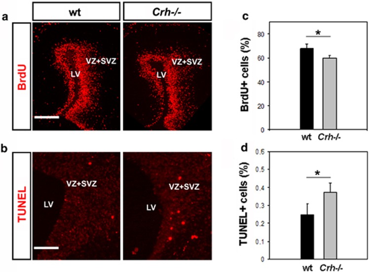
Figure 1.
Altered proliferative and apoptotic properties of neural progenitor cells in the developing brain of Crh−null (Crh−/−) mice. (a) 5-bromo-2-deoxyuridine (BrdU) was administered to the mother at gestational age 14.5 for 2 h. Representative images from brain slices (E14.5) of Crh−/− and wild-type (wt) littermates are shown. Scale bar=20 μm. (c) Graph that shows quantification of the BrdU-positive cell count in 180 × 180 μm2 areas. Data are shown as mean±s.e.m. (n=20 slices of three animals). (b)Representative images from brain slices (E14.5) of Crh−/− and wt littermates after performance of terminal transferase dUTP nick-end labeling (TUNEL) assay. Scale bar=50 μm. (d) Graph that shows quantification of the TUNEL-positive cell count in 180 × 180 μm2 areas. Data are shown as mean±s.e.m. (n=20 slices of four animals) *P<0.05 versus wt mice. vz, svz, LV, lateral ventricle.

