Figure 5.
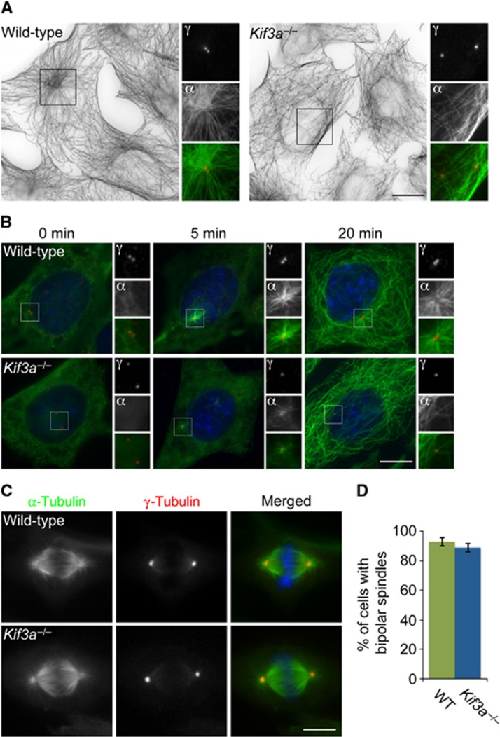
Kif3a is essential for microtubule anchoring at the mother centriole. (A) Immunofluorescence analysis of WT or Kif3a−/− MEFs co-stained for α-tubulin (‘α’, green) and γ-tubulin (‘γ’, red). Large figures are inverted images of α-tubulin staining. The inset shows magnified images of the boxed region. (B) WT and Kif3a−/− MEFs were subjected to a microtubule regrowth assay, fixed at the indicated time points and stained with antibodies to α-tubulin to visualize microtubules (‘α’, green) and γ-tubulin to mark centrosomes (‘γ’, red). (C) Mitotic WT and Kif3a−/− MEFs were stained for α-tubulin and γ-tubulin to visualize mitotic spindles and poles, respectively. (D) Percentage of WT and Kif3a−/− cells with bipolar mitotic spindles. At least 100 cells were analysed per experiment (n=3). Scale bars indicate 5 μm for images in a, 3 μm for images in (B).

