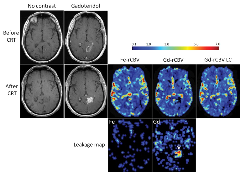
Figure 2:
Axial images of 73-year-old man with GBM show pseudoprogression of disease. T1-weighted MR images without contrast enhancement and with gadoteridol (Gd) obtained before and 3 months after chemoradiotherapy (CRT) show increased contrast enhancement after treatment. Low rCBV (≤1.75) is apparent on parametric maps obtained by using ferumoxytol (Fe-rCBV), gadoteridol (Gd-rCBV), and gadoteridol with leakage correction (Gd-rCBV LC), which indicates pseudoprogression. Leakage map shows absence of contrast extravasation when ferumoxytol (Fe) was used and contrast leakage with gadoteridol (arrow).

