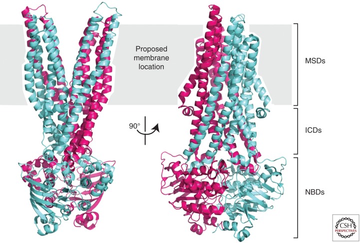
Figure 2.
Ribbon diagram representation of the outward-facing conformation of Sav1866 in two orthogonal views. The subunits of the dimer are shown in blue and purple. Note that the two transmembrane helical bundles move apart as they near the extracellular side of the membrane (PDB 2HYD [Dawson and Locher 2006]).

