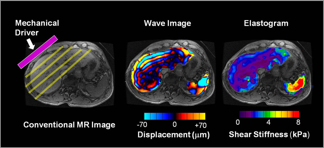
Fig 1.
MRE is performed in three steps. (a) A source of vibration is placed on the surface of the body to generate mechanical waves in the tissues of interest. (b) A special MRE pulse sequence with synchronized motion encoding gradients is used to image the micron-level cyclic displacements caused by the propagating waves. The wavelength of the shear waves is longer in stiffer tissues and shorter in softer tissues. (c) The wave images are then automatically processed with an “inversion algorithm” to create quantitative images depicting the stiffness of tissue. (For these illustrations, the portions of the wave image and elastogram corresponding to liver and spleen have been superimposed on a conventional MR image.)

