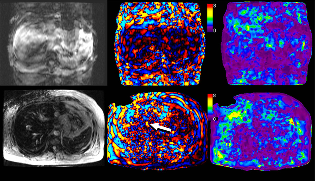
Fig 17.
Situations in which MRE exams may fail. Top row shows severe breath hold artifacts degrading the MRE study in a patient who could not hold breath consistently. Note that waves are not well seen in the wave image and therefore the elastogram (c) is not reliable. Bottom row shows an MRE study in a patient with hemochromatosis and high iron content. The lack of signal from the liver prevents the mechanical waves from being visualized. Without adequate wave data, the elastogram provided by the inversion algorithm is not valid.

