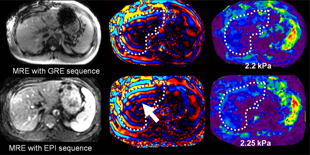
Fig 18.
Patient with moderate hemochromatosis, controlled with multiple phlebotomies. In the top row, MRE was performed with a GRE based technique. Fortunately, the hepatic signal was sufficient to obtain adequate visualization of shear waves, resulting in a valid elastogram. The bottom row shows a follow-up MRE acquisition performed with a spin-echo based EPI technique, designed to be less affected by hepatic iron. The magnitude image demonstrates increased relative signal in the liver, providing improved depiction of waves (arrow). The elastogram provided similar values to the GRE-based measurement, but with improved confidence.

