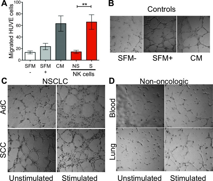
Figure 5.
Proangiogenic activity potential of NK cells. (A) Analysis of the capacity of supernatants from NSCLC-derived NK cells to induce endothelial cell chemotaxis. SFM-, serum-free medium as a negative control, containing RPMI 1640 alone. SFM+, serum-free RPMI 1640 with 1% l-glutamine, fibroblast growth factors (10 ng of acidic fibroblast growth factor plus 10 ng of basic fibroblast growth factor/ml), epidermal growth factor (10 ng/ml), heparin (0.1 mg/ml), and hydrocortisone (010 µg/ml). CM, complete medium containing SFM+ medium supplemented with 10% FBS as a positive control. Supernatants were isolated from AdC and SCC NK cells cultured for 6 hours either unstimulated (NS) or stimulated (S) with PMA and ionomycin. Supernatants from stimulated NK cells showed enhanced induction of chemotaxis compared to supernatants from unstimulated NK cells (NS). Similar data were obtained from AdC and SCC supernatants individually. Media containing only PMA and ionomycin had little chemotactic activity (data not shown). **P < .01. (B–D) NK cells from patients with NSCLC promote endothelial cell capillary-like morphogenesis, (B) positive (SFM+; CM) and negative (SFM-) controls as above. (C) Supernatants from NK cells isolated from AdCs that were stimulated for 6 hours with PMA and ionomycin (stimulated) showed induction of endothelial cell morphogenesis compared to supernatants from unstimulated NK cells. Supernatants from NK cells isolated from SCC showed induction of morphogenesis even when unstimulated, which was further enhanced upon stimulation, indicating that these cells harbor a strong angiogenic activity. (D) NK cells isolated from the peripheral blood (blood) or lung tissues (lung) of non-oncologic patients did not show significant enhancement of morphogenesis in the presence or absence of stimulation.

