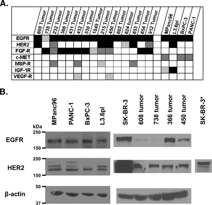
Figure 1.
(A) Relative activation of selected RTKs for 15 patient-derived pancreatic cancer lysates and four established pancreatic cancer cell lines (black, more than three times the threshold; dark gray, two to three times the threshold; light gray, one to two times the threshold). (B) Western blots for EGFR and HER2 in four patient-derived pancreatic cancers, established pancreatic cancer cell lines, and SK-BR-3, a breast cancer cell line (asterisk represents a 1-s exposure).

