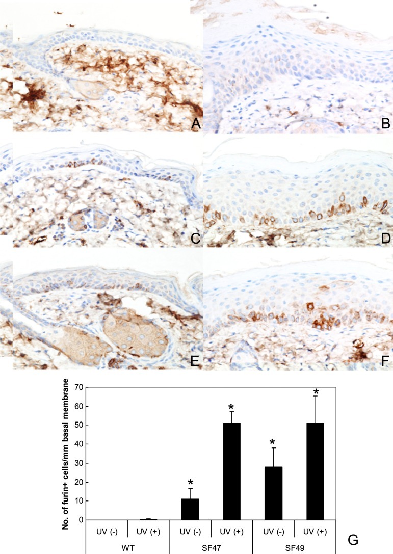
Figure 2.
Immunohistochemistry of furin in dorsal mouse epidermis. Panels A (WT SKH-1), C (F47), and E (F49) depict control unirradiated epidermis. Panels B (WT), D (SF47), and F (SF49) show UV-irradiated epidermis. Both the unirradiated (A) and irradiated WT epidermis (B) show negative or marginal immunostain. Panel C (SF47) exhibits several furin-positive basal keratinocytes that further increased together with occasional positive parabasal keratinocytes after UV irradiation (D). Epidermis from SF49 (E) shows increased expression of furin in basal keratinocytes after UV exposure (F). Furin IHC and hematoxylin counterstain x 150. The histogram (G) shows an increase in the number of positive basal keratinocytes in transgenic epidermis. Asterisks indicate significantly different changes when compared with the respective WT (P < .001).

